Order In Which Light Passes Through The Eye Quizlet
Breaking News Today
Mar 18, 2025 · 6 min read
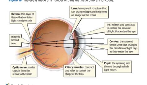
Table of Contents
The Order in Which Light Passes Through the Eye: A Comprehensive Guide
Understanding how light travels through the eye is fundamental to comprehending vision. This intricate process involves a series of structures, each playing a crucial role in focusing light onto the retina and ultimately translating it into the images we see. This comprehensive guide will detail the order in which light passes through the eye, clarifying the function of each component along the way. We'll also explore common misconceptions and offer tips for improving your understanding of this fascinating biological mechanism.
Keywords: eye anatomy, light pathway, cornea, pupil, lens, retina, vision, optics, physiology, ophthalmology, visual perception.
1. The Cornea: The Eye's First Line of Defense
The journey of light begins at the cornea, the eye's transparent outer layer. This dome-shaped structure is responsible for the initial refraction – the bending – of light rays. Because of its curved shape and refractive index, the cornea significantly contributes to focusing the light onto the retina. The cornea's smooth, curved surface is crucial; any irregularities can lead to blurry vision (as seen in conditions like keratoconus). The cornea also acts as a protective barrier, shielding the delicate inner structures of the eye from dust, debris, and microorganisms. Its highly sensitive nerve endings contribute to the blink reflex, a vital protective mechanism.
2. The Aqueous Humor: Maintaining Intraocular Pressure
After passing through the cornea, light enters the aqueous humor, a clear, watery fluid that fills the anterior chamber of the eye – the space between the cornea and the lens. This fluid plays several important roles. It maintains the intraocular pressure (IOP), preventing the eyeball from collapsing. This pressure is crucial for maintaining the shape and function of the eye. The aqueous humor also nourishes the cornea and lens, providing essential nutrients and removing waste products. Imbalances in aqueous humor production and drainage can lead to glaucoma, a condition characterized by elevated IOP and potential damage to the optic nerve.
3. The Pupil and Iris: Regulating Light Intake
Next, light passes through the pupil, the black circular opening at the center of the iris. The iris, the colored part of the eye, acts as a diaphragm, controlling the size of the pupil. In bright light, the iris constricts, making the pupil smaller to reduce the amount of light entering the eye. In dim light, the iris dilates, widening the pupil to allow more light to reach the retina. This dynamic adjustment of pupil size is crucial for adapting to varying light conditions and maintaining optimal visual acuity. The iris's muscles, controlled by the autonomic nervous system, perform this intricate regulation seamlessly.
4. The Lens: Fine-Tuning the Focus
After traversing the pupil, light reaches the lens, a transparent, biconvex structure located behind the iris. The lens plays a crucial role in focusing light onto the retina. Unlike the cornea, which provides a fixed amount of refraction, the lens's shape can be altered by tiny muscles called ciliary muscles. This process, known as accommodation, allows the eye to focus on objects at different distances. For near vision, the ciliary muscles contract, making the lens more rounded and increasing its refractive power. For distant vision, the ciliary muscles relax, allowing the lens to flatten and reduce its refractive power. The ability to accommodate diminishes with age, a phenomenon known as presbyopia, leading to difficulty focusing on near objects.
5. The Vitreous Humor: Supporting the Eye's Shape
Light then passes through the vitreous humor, a clear, gel-like substance that fills the posterior chamber of the eye – the space between the lens and the retina. This large volume of vitreous humor maintains the shape of the eyeball and helps to keep the retina in place. The vitreous humor also transmits light to the retina with minimal scattering. As we age, the vitreous humor can shrink and detach from the retina, potentially leading to floaters or retinal tears.
6. The Retina: Translating Light into Signals
Finally, light reaches the retina, the light-sensitive inner lining of the eye. The retina contains millions of photoreceptor cells – rods and cones – that convert light energy into electrical signals. Rods are responsible for vision in low light conditions, while cones are responsible for color vision and sharp vision in bright light. These signals are then transmitted through the optic nerve to the brain, where they are interpreted as images. The macula, a small region in the center of the retina, contains a high concentration of cones and is responsible for central, high-resolution vision. The optic disc, also known as the blind spot, is the area where the optic nerve exits the eye, lacking photoreceptor cells.
Common Misconceptions about the Order of Light Passage
A common misconception revolves around the order of the pupil and lens. Some mistakenly believe the light passes through the lens before the pupil. However, the pupil acts as a gatekeeper after the light has passed through the cornea and aqueous humor, controlling the amount of light that reaches the lens.
Another common error involves overlooking the vitreous humor's role. While the cornea, lens, and retina are often highlighted, the importance of the vitreous humor in maintaining the eye's shape and transmitting light is often understated.
Finally, the role of accommodation and its relation to the lens is frequently misunderstood. Many fail to grasp the dynamic nature of lens shape change, essential for focusing on objects at varying distances.
Improving Your Understanding: Interactive Learning
To solidify your understanding of this intricate process, consider these interactive approaches:
- Visual Aids: Use diagrams, animations, and videos to visualize the pathway of light. Many online resources offer detailed illustrations and interactive models of the eye.
- Quizzes and Flashcards: Regularly test your knowledge using online quizzes and flashcards. This repeated engagement will reinforce your learning.
- Real-world Applications: Relate the light pathway to common eye conditions, such as nearsightedness (myopia), farsightedness (hyperopia), astigmatism, and cataracts. Understanding how these conditions affect the pathway of light deepens comprehension.
Conclusion: A Journey of Light and Vision
The order in which light passes through the eye is a testament to the remarkable complexity and efficiency of the visual system. From the initial refraction by the cornea to the intricate processing by the retina, each structure plays a vital role in transforming light into the images we perceive. Understanding this process provides a valuable foundation for appreciating the marvel of vision and the delicate mechanisms that enable us to experience the world around us. By actively engaging with visual aids, interactive learning tools, and relating the concepts to real-world applications, you can achieve a thorough understanding of the fascinating journey of light through the eye. This knowledge is not only academically enriching but also crucial for appreciating the importance of maintaining ocular health.
Latest Posts
Latest Posts
-
Which Best Describes The Terrorist Planning Cycle
Mar 18, 2025
-
Cdl Combination Test Questions And Answers Pdf
Mar 18, 2025
-
Life Insurance Exam Questions And Answers Pdf
Mar 18, 2025
-
The Direct Carry Is Used To Transfer A Patient
Mar 18, 2025
-
The Emancipation Proclamation Of January 1 1863 Quizlet
Mar 18, 2025
Related Post
Thank you for visiting our website which covers about Order In Which Light Passes Through The Eye Quizlet . We hope the information provided has been useful to you. Feel free to contact us if you have any questions or need further assistance. See you next time and don't miss to bookmark.
