The Small Space Between Neurons Is Called ____________________.
Breaking News Today
Mar 15, 2025 · 6 min read
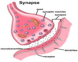
Table of Contents
The Synaptic Cleft: The Tiny Gap That Bridges Neural Communication
The small space between neurons is called the synaptic cleft. This minuscule gap, measuring only about 20-40 nanometers wide, is a crucial component of the nervous system, acting as a bridge for communication between neurons, and ultimately, controlling everything from our thoughts and movements to our emotions and sensations. Understanding the synaptic cleft and the intricate processes that occur within it is essential to comprehending the complexities of the brain and nervous system function, as well as neurological disorders.
What Happens at the Synapse?
The synapse, encompassing the presynaptic terminal, the synaptic cleft, and the postsynaptic membrane, is the site of neuronal communication. This communication, known as synaptic transmission, is not a direct electrical connection but rather a sophisticated chemical process. The arrival of an electrical signal, or action potential, at the presynaptic terminal triggers a series of events:
-
Neurotransmitter Release: The action potential causes voltage-gated calcium channels to open in the presynaptic terminal. This influx of calcium ions initiates the fusion of synaptic vesicles, small membrane-bound sacs containing neurotransmitters, with the presynaptic membrane. The neurotransmitters are then released into the synaptic cleft via exocytosis.
-
Diffusion Across the Synaptic Cleft: Once released, neurotransmitters diffuse across the tiny gap of the synaptic cleft. This diffusion is a passive process, driven by the concentration gradient of the neurotransmitter. The small distance of the cleft ensures that this diffusion process happens relatively quickly, facilitating rapid communication between neurons.
-
Receptor Binding: On the other side of the cleft, the postsynaptic membrane contains receptors specific to the released neurotransmitters. These receptors are proteins embedded in the membrane, and their binding with the neurotransmitter initiates a change in the postsynaptic neuron. This change can be either excitatory, increasing the likelihood of the postsynaptic neuron firing an action potential, or inhibitory, decreasing the likelihood.
-
Signal Termination: The neurotransmitter's action doesn't persist indefinitely. Several mechanisms ensure the termination of the signal, including enzymatic degradation (breakdown of the neurotransmitter by enzymes in the synaptic cleft), reuptake (reabsorption of the neurotransmitter by the presynaptic terminal), and diffusion away from the synapse.
The Importance of the Synaptic Cleft's Dimensions
The precise dimensions of the synaptic cleft are critical for efficient synaptic transmission. If the cleft were significantly wider, the diffusion of neurotransmitters would be slower, leading to a delay in neuronal communication. This could have significant consequences, impairing rapid responses to stimuli and impacting cognitive function. Conversely, a much narrower cleft might lead to uncontrolled neurotransmitter spillover and unwanted cross-talk between synapses. The current dimensions optimize the speed and fidelity of neurotransmission.
Types of Synapses
Synapses are not all the same. They are classified based on various factors, including:
-
Electrical vs. Chemical Synapses: The majority of synapses in the nervous system are chemical synapses, as described above. However, electrical synapses exist, characterized by direct electrical coupling between neurons through gap junctions. These gap junctions allow for rapid, bidirectional transmission of electrical signals, but they lack the fine-tuning and plasticity of chemical synapses.
-
Axodendritic, Axosomatic, and Axoaxonic Synapses: These classifications refer to the location of the synapse on the postsynaptic neuron. Axodendritic synapses are formed between the axon terminal of one neuron and the dendrite of another. Axosomatic synapses connect the axon terminal to the soma (cell body) of the postsynaptic neuron. Axoaxonic synapses occur between two axons. These different synaptic locations can influence the strength and impact of synaptic transmission.
-
Excitatory vs. Inhibitory Synapses: As mentioned earlier, synapses can be either excitatory or inhibitory, depending on the type of neurotransmitter released and the receptors present on the postsynaptic membrane. Excitatory synapses promote the firing of action potentials in the postsynaptic neuron, while inhibitory synapses suppress it. This balance between excitation and inhibition is crucial for maintaining normal nervous system function.
The Synaptic Cleft and Neurological Disorders
Disruptions in synaptic transmission, often involving problems within the synaptic cleft, are implicated in a wide array of neurological and psychiatric disorders. These disruptions can arise from various factors:
-
Neurotransmitter Imbalances: Conditions like depression and anxiety are often associated with imbalances in neurotransmitter levels in the synaptic cleft. For example, a deficiency in serotonin or dopamine can lead to impaired synaptic transmission and contribute to depressive symptoms.
-
Receptor Dysfunction: Many neurological disorders involve abnormalities in the receptors located on the postsynaptic membrane. These abnormalities can affect the binding of neurotransmitters, leading to altered signal transduction and contributing to symptoms of the disorder.
-
Autoimmune Diseases: Certain autoimmune diseases can target components of the synapse, causing inflammation and damage to the synaptic cleft and impairing communication between neurons.
-
Neurodegenerative Diseases: In neurodegenerative diseases like Alzheimer's and Parkinson's disease, the degeneration of neurons leads to a progressive loss of synapses and impaired synaptic transmission. This synaptic dysfunction contributes to the cognitive decline and motor impairments characteristic of these diseases.
Specific examples of neurological disorders linked to synaptic dysfunction include:
-
Alzheimer's Disease: Characterized by amyloid plaques and neurofibrillary tangles that disrupt synaptic function, leading to memory loss and cognitive decline.
-
Parkinson's Disease: Involves the degeneration of dopamine-producing neurons in the substantia nigra, resulting in a deficiency of dopamine in the synaptic cleft and causing motor impairments.
-
Schizophrenia: Associated with disruptions in the dopaminergic and glutamatergic systems, affecting synaptic transmission and contributing to psychotic symptoms.
-
Epilepsy: Characterized by excessive neuronal activity, often due to imbalances in excitatory and inhibitory synaptic transmission.
-
Multiple Sclerosis (MS): An autoimmune disease that attacks the myelin sheath surrounding nerve fibers, disrupting nerve impulse transmission and potentially impacting synaptic function.
Therapeutic Interventions Targeting the Synapse
Many therapeutic interventions for neurological and psychiatric disorders aim to modulate synaptic transmission within the synaptic cleft. These interventions include:
-
Pharmacological Agents: Many drugs act by influencing neurotransmitter levels, receptor activity, or reuptake mechanisms within the synapse. Antidepressants, for example, often increase serotonin levels in the synaptic cleft. Antipsychotics may block dopamine receptors.
-
Deep Brain Stimulation (DBS): A surgical procedure involving the implantation of electrodes into specific brain regions to modulate neuronal activity and potentially improve synaptic function. DBS is used for conditions like Parkinson's disease and essential tremor.
-
Non-Invasive Brain Stimulation Techniques: Techniques like transcranial magnetic stimulation (TMS) and transcranial direct current stimulation (tDCS) use magnetic or electrical fields to modulate neuronal activity and potentially improve synaptic plasticity.
Ongoing Research and Future Directions
Research on the synapse continues to advance our understanding of its complex mechanisms and its role in health and disease. Scientists are investigating new therapeutic targets within the synapse, exploring novel ways to modulate synaptic transmission and potentially develop more effective treatments for neurological and psychiatric disorders. Advanced imaging techniques are providing unprecedented insights into the structure and function of synapses in living organisms. This ongoing research promises to further elucidate the intricacies of this tiny gap that plays such a massive role in our lives. By understanding the complex processes at play within the synaptic cleft, we move closer to creating effective therapies for a vast range of debilitating conditions.
The synaptic cleft, while incredibly small, is a pivotal site for the intricate communication that underpins all aspects of our nervous system function. Its precise dimensions, the dynamic interplay of neurotransmitters and receptors, and its susceptibility to dysfunction highlight its crucial role in both health and disease. Future research into this fascinating and vital area holds immense potential for improving human health.
Latest Posts
Latest Posts
-
According To The Theory Of Plate Tectonics
Mar 15, 2025
-
Which Of These Events Would Be A Result Of Inflation
Mar 15, 2025
-
Receptors For Nonsteroid Hormones Are Located In
Mar 15, 2025
-
Dead In 5 Seconds Was Dedicated To
Mar 15, 2025
-
Which Of The Following Do Not Contribute To Tension Headaches
Mar 15, 2025
Related Post
Thank you for visiting our website which covers about The Small Space Between Neurons Is Called ____________________. . We hope the information provided has been useful to you. Feel free to contact us if you have any questions or need further assistance. See you next time and don't miss to bookmark.
