Identify The Parts Of The Fibrous Layer
Breaking News Today
Mar 28, 2025 · 6 min read
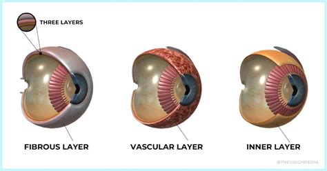
Table of Contents
Identify the Parts of the Fibrous Layer: A Deep Dive into the Sclera and Cornea
The fibrous layer, also known as the tunica fibrosa, is the outermost layer of the eyeball. It's a tough, protective layer crucial for maintaining the eye's shape and providing structural support. Understanding its components is essential for comprehending ocular anatomy and various ophthalmological conditions. This comprehensive guide will delve into the intricate details of the fibrous layer, exploring its two main parts: the sclera and the cornea.
The Sclera: The White of the Eye
The sclera, commonly known as the "white of the eye," forms the posterior five-sixths of the fibrous layer. Its primary function is to protect the delicate inner structures of the eye, including the retina, choroid, and vitreous humor. Imagine it as a strong, protective shell safeguarding the eye's precious contents.
Composition and Structure of the Sclera
The sclera is composed of dense, irregular connective tissue primarily consisting of:
-
Collagen fibers: These provide the sclera's strength and resilience, resisting the internal pressure of the eye (intraocular pressure). The intricate arrangement of these fibers gives the sclera its characteristic white opaque appearance.
-
Elastic fibers: These fibers contribute to the sclera's flexibility and ability to withstand minor deformations without damage. They help maintain the eye's shape and prevent significant stretching.
-
Fibroblasts: These are the cells responsible for producing and maintaining the collagen and elastic fibers of the sclera. Their activity is vital for the sclera's structural integrity and repair processes.
The sclera's structure isn't uniform throughout. It's thicker posteriorly, providing greater protection at the back of the eye where it's more vulnerable to impact. Anteriorly, it becomes progressively thinner, merging with the cornea at the limbus. This transition zone is important for the proper functioning of both structures.
Scleral Perforations and Passages
Despite its robust nature, the sclera has several strategic openings that allow passage for critical structures:
-
Posterior scleral foramina: These tiny openings allow the optic nerve to exit the eye, connecting the retina to the brain. The surrounding tissue is highly vascularized and supportive to the optic nerve.
-
Anterior scleral foramina: These openings accommodate the passage of the extraocular muscles, crucial for controlling eye movement. These muscles attach to the sclera allowing for the precise and coordinated movements we rely on for clear vision.
-
Episcleral vessels: A network of blood vessels lies between the sclera and the conjunctiva (the thin membrane covering the sclera and inner eyelid). These vessels provide oxygen and nutrients to the sclera's outer layers.
Clinical Significance of Scleral Disorders
Several pathological conditions can affect the sclera:
-
Scleritis: This is an inflammation of the sclera, often associated with autoimmune diseases. It can present with pain, redness, and impaired vision. Early diagnosis and treatment are crucial to prevent serious complications.
-
Episcleritis: This is a milder inflammation of the episclera, the thin layer of tissue covering the sclera. It usually presents with localized redness and discomfort, often resolving spontaneously.
-
Scleral thinning: This can occur due to various factors, including age-related degeneration or certain medical conditions. It can lead to increased intraocular pressure and potentially serious vision problems.
-
Scleral rupture: A serious injury to the eye can cause a scleral rupture, potentially leading to the loss of vitreous humor or retinal detachment. Immediate medical attention is required.
The Cornea: The Eye's Transparent Window
The cornea, comprising the anterior sixth of the fibrous layer, is a transparent, avascular structure that plays a critical role in focusing light onto the retina. Unlike the sclera, the cornea's transparency is essential for clear vision. Its unique structure and composition contribute to this critical function.
Composition and Structure of the Cornea
The cornea is composed of five distinct layers:
-
Epithelium: This outermost layer is a stratified squamous epithelium, constantly regenerating and protecting the underlying structures from abrasion and infection. Its rapid healing capacity is essential for maintaining corneal integrity.
-
Bowman's layer: A thin, acellular layer providing structural support and acting as a barrier against infections. It does not regenerate if damaged.
-
Stroma: This is the thickest layer of the cornea, consisting of highly organized collagen fibrils and keratocytes (corneal fibroblasts). The precise arrangement of these collagen fibers is crucial for corneal transparency. Any disruption in this organization can lead to corneal opacity.
-
Descemet's membrane: A basement membrane separating the stroma from the endothelium. This layer is remarkably resistant to damage and plays a significant role in maintaining corneal hydration.
-
Endothelium: This innermost layer is a monolayer of specialized cells that maintain corneal hydration by pumping fluid out of the stroma. The endothelium's function is crucial for preventing corneal swelling and preserving its transparency. Its ability to regenerate is limited, making it particularly vulnerable to damage.
Corneal Transparency and its Mechanism
The cornea's transparency is achieved through several factors:
-
Regular arrangement of collagen fibers: The precisely organized collagen fibrils minimize light scattering, ensuring clear transmission of light to the retina.
-
Avascular nature: The cornea's lack of blood vessels prevents light scattering by blood cells. Nutrients are obtained through diffusion from the surrounding tissues and aqueous humor.
-
Hydration level: The cornea's hydration level is meticulously controlled by the endothelium, preventing swelling that could compromise its transparency.
Clinical Significance of Corneal Disorders
The cornea is susceptible to various diseases and injuries, many of which can severely impact vision:
-
Corneal ulcers: Infections or injuries can lead to corneal ulcers, potentially causing scarring and vision loss. Prompt treatment with antibiotics or antiviral medications is essential.
-
Keratitis: Inflammation of the cornea, often caused by infections or autoimmune disorders. Symptoms can include pain, redness, blurred vision, and light sensitivity.
-
Corneal dystrophies: These are inherited disorders affecting the cornea's structure and transparency, potentially leading to visual impairment.
-
Corneal edema: Swelling of the cornea due to impaired endothelial function or other factors. This can lead to blurred vision and discomfort.
-
Corneal trauma: Injuries to the cornea, such as scratches or lacerations, can cause significant damage, potentially leading to infection and scarring.
The Limbus: The Transition Zone
The limbus is the crucial transition zone where the cornea and sclera meet. This area is highly specialized and plays several important roles:
-
Connective tissue transition: The limbus marks the transition between the organized collagen fibers of the cornea and the more irregularly arranged fibers of the sclera.
-
Stem cell niche: The limbus contains limbal stem cells, crucial for the regeneration of the corneal epithelium. Damage to the limbal stem cells can lead to significant corneal problems.
-
Vascularization: The limbus is highly vascularized, providing nutrients and oxygen to the avascular cornea.
-
Immune surveillance: The limbus contains immune cells that play a vital role in protecting the cornea from infection.
Conclusion: The Fibrous Layer's Crucial Role in Vision
The fibrous layer, encompassing the sclera and cornea, is a remarkable structure crucial for maintaining the eye's structural integrity and facilitating clear vision. Its components are highly specialized, each contributing to its overall function. Understanding the intricate details of the fibrous layer, its various components, and the associated clinical significance is essential for any healthcare professional involved in eye care. Further research into the complexities of this layer continues to unveil new insights into maintaining healthy vision and treating ocular diseases. The intricacies of the sclera and cornea highlight the remarkable engineering of the human eye, a testament to the power of nature's design. The synergistic relationship between these two components ensures the eye's overall protection and visual acuity, underscoring their vital roles in human vision.
Latest Posts
Latest Posts
-
The Division Of The Cytoplasm Is Called
Mar 30, 2025
-
Comedy Is To Laughter As Insecurity Is To
Mar 30, 2025
-
Which Of The Following Poses A Physical Security Risk
Mar 30, 2025
-
Which One Of These Is An Amino Group
Mar 30, 2025
-
Which Of The Following Statements Is True About Managerial Compensation
Mar 30, 2025
Related Post
Thank you for visiting our website which covers about Identify The Parts Of The Fibrous Layer . We hope the information provided has been useful to you. Feel free to contact us if you have any questions or need further assistance. See you next time and don't miss to bookmark.
